
VegFizz Hydrogen Scam
September 6, 2022
Part 1 How Ultraviolet Radiation Damages the Skin and Causes Photoaging
October 24, 2022NREM and REM Sleep 101

The Behavior of Sleep
My PhD advisor frequently reminded me that the primary consequence of inadequate sleep is sleepiness. And while sleep is principally understood through the subjective consequences of its absence, simply “feeling tired” does not necessarily provide useful insights into the specific way a persons’ sleep might be impaired nor necessarily reflect healthy or disordered sleep. You know what sleep is – through experience you understand it and in its absence, you appreciate how important it is. Sleepiness and sleep are so ubiquitous and ordinary that it is only in our third blog installation on sleep that I will offer a formal definition: Sleep is “a reversible behavioral state of perceptual disengagement from, and unresponsiveness to, the environment” – Carskadon and Dement (2011)
Sleep is not some unitary state of unconsciousness, but a complex behavioral state, organized into robust cycles of two major sleep subtypes: rapid eye movement (REM) sleep and non-rapid eye movement (NREM) sleep. Recent advances in neuroscience and sleep research have emphasized the importance of transition states, too, further illustrating the complexity of sleep behavior. These sleep stages have behavioral and electrophysiological features that distinguish them; and each of them are produced and maintained by the activity of distinct brain regions and specific neurochemical environments.
Non-Rapid Eye Movement (NREM) Sleep
NREM sleep is characterized by: (i) recumbent posture and decreased motor activity (without atonia); (ii) absence of sensation and perception, with increased threshold for arousal; and (iii) loss of consciousness. NREM can be divided into NREM Stage 1, NREM Stage 2, and NREM Stage 3 (and NREM Stage 4, depending on who you ask). NREM Stage 3 is also known as “deep sleep” or “slow wave sleep” (SWS) as cortical neurons are highly synchronized in their up- and down-states during this stage, underlying the low frequency (0-4Hz), high amplitude signal in EEG that typifies it. This “deepest” substage of NREM is thought to be essential for the homeostatic regulation of sleep and wake (for more, check out our earlier blog here). The specific electrophysical and neurochemical environment of NREM sleep is associated with several important biological functions, including the pulsed release of growth hormone and testosterone in males [1] and cognition. NREM sleep is associated with the consolidation and preservation of episodic memories [2] – importantly, it appears that there are specific patterns of cortical reactivation (which dominate, but are not exclusive to, NREM sleep) that are intrinsically tied to the reinforcement or creation of associations (aka learning), while other waveforms (which are reflections of underlying patterns of cortical connectivity) appear to play a role in the weakening of memories [3]. Advances in the fields of optogenetics and electrophysiology have begun to demonstrate that even within specific substages of NREM sleep, for example, there is incredible diversity in biological conditions and these conditions are important for specific processes once assumed to be categorically associated with an entire stage of sleep.
Rapid Eye Movement (REM) Sleep
Behaviorally, REM sleep is characterized by a combination of: (i) lack of muscle tone from spinal motor neuron inhibition or “muscle atonia”; (ii) vivid, internally generated and often bizarre/illogical sensory perception or “dreaming”; and (iii) rapid conjugate eye movement, thought to reflect dreaming. Total nightly concentration of REM sleep will change dramatically throughout life. At birth, 40% of infant sleep is dominated by REM sleep, with that decreasing to 20-25% by year 10 and 15% by middle adulthood [4,5]. Numerous reports suggest that REM sleep may be essential to brain development, and there are ongoing efforts to understand how the neurochemical environment and patterns of cortical- and sub-cortical connectivity associated with REM sleep might contribute to early development across species. One note here is that a hallmark of early brain development is rapid, widespread overproduction of new connections among cells, called synapses [6,7]. These synapses are the physical substrate upon which connective associations between cells are maintained. For the sake of maintaining selective, useful associations between cells (rather than having all cells be similarly associated with one another), this overproduction of synapses necessitates the subsequent widespread strategic pruning (removal or downscaling) of these formations. Over the last several years, numerous reports have demonstrated a role for REM sleep in the reorganization of memories – specifically, it seems REM sleep is essential (or, at least, permissive) to the weakening of associations (aka synaptic pruning) [8-10].
While REM does appear to be principally organized by pontine nuclei in the brainstem, the behavioral features of REM (rapid eye movement, dreaming, muscle atonia) and other physiological changes associated with REM (increased theta frequency oscillations in the cortex and hippocampus, PGO waves) appear to be maintained by distinct neural systems. For example, inhibition of muscle tone during REM sleep emerges from parallel inhibitory GABAergic projections from the sublaterodorsal nucleus (SLD) of the tegmentum to spinal motor neurons, while the hippocampal theta oscillations that appear to be an important part of cognition are driven by glutamatergic signal from the pontine reticular formation to the basal forebrain [11]. There are also instances of “phasic REM sleep”, during which individuals may experience bursts of muscle twitches, and we now appreciate that the transitions from non-REM sleep into REM sleep – known as transition REM (tREM) or the intermediate state (IS) may play a specialized role in cognition [12,13].
Polysomnography
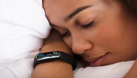
In their effort to unpack the complexity of sleep and sleep loss, scientists (and interested consumers) can use a variety of tools: sleep surveys, actigraphy, or next-generation wearables (Oura, FitBit, Apple Watch), and mobile health applications. However, for the purposes of diagnosing and monitoring clinical sleep disorders, sleep apnea, and seizure disorders, clinicians rely on polysomnography (PSG). PSG is a multi-parametric test used in the study of sleep and the diagnosis of sleep disorders – its name is derived from both Greek (polus for “many”; graphein for “write”) and Latin (somnus for “sleep”). Polysomnography typically combines electroencephalography (EEG) with electromyography (EMG) and, in the case of human investigations, electromyography (EOG) [14].
EEG is a non-invasive method for recording electrical activity from the scalp, giving researchers and clinicians valuable information about the activity of the cortex, the outermost layer of the brain. EEG offers very good temporal resolution (sampling rates are typically between 200-2000Hz), and variable spatial resolution that depends on the number of electrodes used. EEG enables the identification of cortical oscillations which reflect the underlying synchronization and connectivity of the cortex. It is an essential part of PSG but is not sufficient on its own for the investigation of sleep. EOG is a tool for measuring the electrical activity of the eyes (which are dipoles), enabling the assessment of eye movements. Because cortical activity during REM sleep is similar to waking and because rapid conjugate eye movements are a hallmark of REM sleep, the use of EOG for the measurement of eye movements is an essential complement to EEG in polysomnography for humans. EMG is a technique for recording electrical activity from skeletal muscle. The use of EMG enables researchers to identify periods of movement (and stillness). Because muscle is a hallmark of REM sleep, EMG is used in conjunction with EEG and EOG in humans for reliable identification of REM. As EOG is impractical in many model organisms like rats and mice, polysomnography in these species is usually a combination of EEG and EMG only.
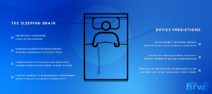
So what is the point of all of this? Well, consider that one substage of sleep has numerous features, each of which are regulated by dynamic interactions across multiple brain regions. So, when a sleep tracking device says that you should pay attention to your REM sleep, what does that mean? What should you do with that information? Several complex processes underlie each part of sleep, so it is difficult to know what (if anything) can/should be done if your sleep behavior varies from the “norm”, especially with so much normal variability between individuals. Wearables track movement to infer/predict periods of muscle tone associated with REM sleep, but what if you are a person that naturally gets more phasic REM sleep, characterized by bursts of muscle movement?** Similarly, you could be still all night long, and have periods of NREM sleep mistaken for REM, or even have deficits in the projections from the basal forebrain to the hippocampus that, during the earlier stages of REM sleep each night, are important for the integration of recently formed associations. To Oura’s credit, they use multiple endpoints to make probabilistic decisions about a person’s sleep staging – and they do a pretty good job of it [15,16]. Despite their success, it remains terribly difficult for consumers (and patients, for that matter) to judge the “quality of their sleep” because many important elements of this massively complex behavior cannot reasonably be assessed in parallel outside of the sleep clinic nor in real time. In our next installation, we’ll do a deep dive into the efficacy of different tools for at-home sleep assessment.
** Excessive bursts of muscle movement, or inability to inhibit spinal motor neurons during dreaming (acting
out your dreams) may be a sign of a serious sleep disorder and should be evaluated by an appropriately
credentialed physician in the field of sleep medicine.
1. Assefa, S. Z., Diaz-Abad, M., Wickwire, E. M., & Scharf, S. M. (2015). The functions of sleep. AIMS
Neuroscience, 2(3), 155-171.
2. Stickgold, R. (2009). How do I remember? Let me count the ways. Sleep medicine reviews, 13(5), 305.
3. Kim, J., Gulati, T., & Ganguly, K. (2019). Competing roles of slow oscillations and delta waves in memory
consolidation versus forgetting. Cell, 179(2), 514-526.
4. Davis, K. F., Parker, K. P., & Montgomery, G. L. (2004). Sleep in infants and young children: Part one: normal
sleep. Journal of Pediatric Health Care, 18(2), 65-71.
5. Ehlers, C. L., & Kupfer, D. J. (1989). Effects of age on delta and REM sleep parameters. Electroencephalography
and clinical neurophysiology, 72(2), 118-125.
6. Testa-Silva, G., Loebel, A., Giugliano, M., de Kock, C. P., Mansvelder, H. D., & Meredith, R. M. (2012).
Hyperconnectivity and slow synapses during early development of medial prefrontal cortex in a mouse model for
mental retardation and autism. Cerebral cortex, 22(6), 1333-1342.
7. Innocenti, G. M., & Price, D. J. (2005). Exuberance in the development of cortical networks. Nature Reviews
Neuroscience, 6(12), 955-965.
8. Landmann, N., Kuhn, M., Maier, J. G., Spiegelhalder, K., Baglioni, C., Frase, L., … & Nissen, C. (2015). REM
sleep and memory reorganization: potential relevance for psychiatry and psychotherapy. Neurobiology of learning
and memory, 122, 28-40.
9. Best, J., Diniz Behn, C., Poe, G. R., & Booth, V. (2007). Neuronal models for sleep-wake regulation and synaptic
reorganization in the sleeping hippocampus. Journal of Biological Rhythms, 22(3), 220-232.
10. Aime, M., Calcini, N., Borsa, M., Campelo, T., Rusterholz, T., Sattin, A., … & Adamantidis, A. (2022). Paradoxical
somatodendritic decoupling supports cortical plasticity during REM sleep. Science, 376(6594), 724-730.
11. Scammell, T. E., Arrigoni, E., & Lipton, J. O. (2017). Neural circuitry of wakefulness and sleep. Neuron, 93(4),
747-765.
12. Vanderheyden, W. M., George, S. A., Urpa, L., Kehoe, M., Liberzon, I., & Poe, G. R. (2015). Sleep alterations
following exposure to stress predict fear-associated memory impairments in a rodent model of PTSD.
Experimental brain research, 233(8), 2335-2346.
13. Poe, G. R. (2017). Sleep is for forgetting. Journal of Neuroscience, 37(3), 464-473.
14. Van De Water, A. T., Holmes, A., & Hurley, D. A. (2011). Objective measurements of sleep for non‐laboratory
settings as alternatives to polysomnography–a systematic review. Journal of sleep research, 20(1pt2), 183-200.
15. de Zambotti, M., Rosas, L., Colrain, I. M., & Baker, F. C. (2019). The sleep of the ring: comparison of the ŌURA
sleep tracker against polysomnography. Behavioral sleep medicine, 17(2), 124-136.
16. Altini, M., & Kinnunen, H. (2021). The promise of sleep: A multi-sensor approach for accurate sleep stage
detection using the oura ring. Sensors, 21(13), 4302.


